

【The Complete Guide to Spondylolisthesis】Causes, Rehabilitation, and Surgical Timing: Precision Treatment Recommendations from Grade 1 to Grade 5
【Quick Summary】
Spondylolisthesis is a condition in which one vertebra slips forward or backward relative to the vertebra below it, often caused by degeneration, trauma, or a pars fracture. Clinically, it is classified into grades 1 to 5 based on the degree of displacement. Most patients with Grade 1 or Grade 2 spondylolisthesis can improve with physical therapy and core strengthening exercises. However, if symptoms such as neurogenic claudication, cauda equina syndrome develop, or if conservative treatment fails, surgery may need to be considered. Modern medicine now offers precision computer navigation technology, combined with 3D imaging systems and intraoperative O-arm imaging, which enables real-time image guidance during surgery. This allows implant placement errors to be controlled to within 0.1 cm, significantly improving safety—especially for elderly patients and those undergoing complex revision surgeries.
What Is Spondylolisthesis? From Causes to Clinical Grading
Spondylolisthesis is not a single disease but a condition of spinal instability. When the structures that maintain spinal stability—such as the intervertebral discs, facet joints, and ligaments—are damaged, the vertebra can slip out of its normal position, potentially compressing the nerve roots or spinal cord.
1. Common Types of Spondylolisthesis
Based on clinical observations from Dr. Chien-Chun Chang's team, the causes of spondylolisthesis can be classified into five main types::
-
Degenerative:Most commonly seen in women over 50, typically occurring at the L4-L5 level, caused by long-term overuse leading to joint laxity.
-
Isthmic (Pars Defect):More common in adolescent athletes, resulting from a stress fracture of the pars interarticularis, most frequently at L5.
-
Congenital:Caused by abnormal spinal development present at birth.
-
Traumatic: Caused by fracture displacement resulting from severe accidents.
-
Pathologic:Caused by underlying conditions such as tumor or osteoporosis.
2. The Five-Grade Classification System: A Standard for Assessing Severity
Doctors typically evaluate the severity of spondylolisthesis by measuring the percentage of forward slippage of the upper vertebra on lateral X-ray. The vertebra is divided into four equal parts (each representing 25%) , and the degree of slip is classified as follows:
|
Grade |
Slippage Percentage |
Recommended Treatment Direction |
|
Grade I |
< 25% |
Mainly conservative treatment, focusing on core muscle strengthening. |
|
Grade II |
25% - 50% |
Mainly conservative treatment; surgery may be considered if nerve compression is present. |
|
Grade III |
50% - 75% |
Surgical evaluation is usually recommended; strong sense of structural instability. |
|
Grade IV |
75% - 100% |
High risk of nerve damage; surgical correction is recommended. |
|
Spondyloptosis |
> 100% |
Complete vertebral displacement (vertebral drop); highly complex condition requiring care from a specialized team. |
What Are the Symptoms of Spondylolisthesis? Why Do You Feel "Leg Weakness" and "Numbness"?
In the early stages, spondylolisthesis may not cause noticeable symptoms. However, as the slippage progresses, the following three typical signs often appear:
1. Localized Lower Back Pain
Patients often experience worsening pain when standing for long periods, walking, or arching the back backward, while relief is often found when sitting down or leaning forward. This is because the muscles and ligaments around the slipped segment are constantly in a state of compensatory tension.
2. Radiating Leg Pain and Numbness
When the vertebral displacement compresses the nerve root (foramen) , the pain and numbness radiate along the nerve pathway to the buttocks, the back of the thigh, and even down to the calf and sole of the foot. This is commonly known as sciatica.
3. Neurogenic Claudication
This is a hallmark symptom when spinal stenosis coexists with spondylolisthesis. After walking a certain distance, patients feel aching, numbness, and weakness in both legs, forcing them to squat or sit down to rest. Once they rest, the pressure on the nerves temporarily subsides, and they can walk again.
Dr. Chang's Perspective:
Many people mistakenly believe these symptoms are caused by "poor blood circulation," but in fact, they are signs of nerve compression. If spondylolisthesis is not addressed in a timely manner, it can lead to muscle atrophy.
The Golden Window for Conservative Treatment: Can I Avoid Surgery? [1]
For patients with Grade 1 and Grade 2 spondylolisthesis, Dr. Chien-Chun Chang's team prioritizes a systematic conservative treatment approach. The goal is to stabilize the spine through a combination of external support and internal muscle strengthening.
1. Physical Therapy and Core Training
-
Williams' Flexion Exercises:These exercises help reduce lumbar lordosis and relieve stress at the site of the slippage.
-
Deep Core Strengthening:Training the transversus abdominis and multifidus muscles to build a strong muscular corset around the spine.
2. Medication and Lifestyle Adjustments
-
Medication:Non-steroidal anti-inflammatory drugs (NSAIDs) or muscle relaxants may be used to relieve acute pain.
-
Posture Awareness:Avoid bending at the waist when lifting heavy objects, sitting on low stools, or standing/sitting for prolonged periods.
-
Wearing a Brace:A rigid or soft lumbar brace can help limit spinal movement and prevent the slippage from worsening.
3. Pain Management
If oral medications are ineffective, a Transforaminal Epidural Steroid Injection (TFESI) may be considered. This is an advanced type of nerve block that delivers medication precisely to the site of nerve root compression under image guidance, achieving the dual goals of differential diagnosis and targeted pain relief.
The Key Turning Point for Surgery: When Should You Consider It? [2]
When pain becomes severe enough to significantly affect your quality of life, it may be time to consider surgery. Specific indications include:
-
Failed Conservative Treatment:Symptoms continue to worsen despite 3 to 6 months of physical therapy.
-
Neurological Deterioration:Development of foot drop (inability to lift the front of the foot, causing frequent tripping) or noticeable muscle atrophy.
-
Cauda Equina Syndrome:Impaired bowel or bladder function — this is a medical emergency requiring immediate intervention.
-
Progressive Slippage:Imaging studies show a significant increase in the degree of vertebral displacement.

Modern Technology: 3D Computer-Navigated Minimally Invasive Surgery Reduces Risks [3]
In the past, spinal surgery for spondylolisthesis was often viewed by older adults as a "major operation," with concerns about risks such as nerve damage leading to paralysis. However, with advances in technology, we can now utilize the latest surgical equipment—often referred to as the "GPS of spine surgery"—to significantly reduce these risks. The 3D computer navigation system, combined with the O-arm intraoperative imaging system, provides real-time image guidance during surgery.
1. Why Is "Navigation" the Key to Safety?
Traditional surgery relies on 2D X-ray images and the surgeon’s experience. However, in severe cases of spondylolisthesis, the spinal anatomy is often deformed or displaced. The navigation system provides the surgeon with more detailed, real-time images during the procedure, serving as a crucial decision-making aid:
-
Enhanced Accuracy:The navigation imaging system provides real-time 3D images, allowing implant placement to be controlled within 0.1 cm, effectively avoiding critical nerves and blood vessels.
-
Minimally Invasive Incision:Because of the precise localization, there is no need for extensive muscle dissection to visualize the anatomy. The incision is typically only about 2–3 cm, resulting in minimal blood loss.
-
Effective Reduction:With accurate and precise implant placement, the surgeon can effectively realign the displaced vertebra to its proper position.
2. Core Surgical Option: Computer-Navigated Minimally Invasive Lumbar Fusion
For unstable spondylolisthesis, this is the current mainstream surgical approach:
-
Decompression:Removal of hypertrophic ligaments or bone spurs that are compressing the nerves.
-
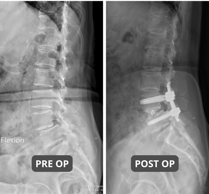
Reduction and Fusion:The slipped vertebra is repositioned to its normal alignment, an interbody cage is placed into the disc space, and screws are precisely inserted under navigation guidance for secure fixation.
-
Advantage:Patients are typically able to get out of bed with the support of a lumbar brace the day after surgery, significantly shortening the hospital stay.
Postoperative Rehabilitation and Long-Term Care for Spondylolisthesis
Surgical success is only the first step. Postoperative "maintenance" is the key to determining how long your spine will last.
-
First 2 Weeks After Surgery:Rest is essential. With the protection of a brace, patients can move and walk freely, but it is advisable to rest more to reduce postoperative soreness and promote wound healing. Strictly follow the principles of "no bending, no prolonged sitting, and no heavy lifting" and wear the rigid back brace correctly.
-
1 Month After Surgery (Healing Phase): Brisk walking on flat ground is permitted to promote fusion between the spinal bone and the implants.
-
3 Months After Surgery (Strengthening Phase):Begin low-impact exercises (such as walking or water walking) and restart core training under the guidance of a physical therapist.
-
Weight Management:For every 1 kg of weight gained, the load on the lumbar spine increases exponentially. Maintaining an ideal BMI is highly recommended.
Conclusion: Precision Medicine Makes "Standing Tall" No Longer a Challenge
Although spondylolisthesis may sound alarming, within the framework of modern medicine, it is a highly manageable and treatable condition. From Grade I preventive rehabilitation to advanced navigated minimally invasive surgery, the key lies in "precise diagnosis" and "timely intervention."
If you or an elderly family member are experiencing chronic low back pain, difficulty walking long distances, or numbness in the legs, be sure to consult a qualified orthopedic surgeon. Confirm the grade of spondylolisthesis through X-ray or MRI imaging.
Spondylolisthesis Surgery FAQ
Q:Is surgery always necessary for spondylolisthesis?
Not necessarily. For patients with mild slippage (Grades I and II) , non-surgical treatment is the first-line option. Most patients can achieve significant improvement through physical therapy, core muscle strengthening, and lifestyle modifications. Surgery is typically recommended only when there is persistent nerve compression, muscle atrophy, neurogenic claudication, or when conservative treatment has failed after three months.
【Key Evidence】
A 2025 comprehensive review published in the Journal of Medical Life stated: "Non-surgical management is the first-line treatment for low-grade spondylolisthesis," with most patients achieving symptom relief through conservative care.[2]
Q:What are the exercise precautions for spondylolisthesis?
Patients with spondylolisthesis should avoid repetitive, excessive lumbar flexion, hyperextension, and sudden twisting movements, as these can place additional stress on an already unstable spinal structure. For example: backbends in yoga, heavy deadlifts, or vigorous swinging motions in badminton or golf—these movements may worsen the degree of slippage.
【Key Evidence】
A 2024 meta-analysis published in the Spine journal confirmed that "lumbar segmental stabilization exercises" significantly improve functional disability in patients with lumbar spondylolisthesis. Movements that involve excessive lumbar extension and sudden twisting should be avoided, as they may aggravate instability at the site of the slippage.[4]
Q:How long does recovery take after minimally invasive surgery?
With 3D computer-navigated minimally invasive surgery, most patients are able to walk within 1–2 days after surgery and are discharged from the hospital within 3–5 days. For desk-based office work, patients can typically return within 2–4 weeks. For jobs that require heavy lifting or manual labor, it is recommended to wait at least 3 months to allow for stable bone fusion before resuming such activities.
【Key Evidence】
1. Recovery Advantages of Navigated Minimally Invasive Surgery
According to a 2024 meta-analysis published in World Neurosurgery, computer-navigated lumbar spine surgery was associated with an average hospital stay reduction of 2.67 days and an average blood loss reduction of 120.62 mL compared to conventional surgery.
2. Evidence on Return-to-Work Outcomes
According to a 2022 study published in Spine journal, among patients who underwent minimally invasive lumbar fusion, 85.7% returned to work within 30 days; among those who underwent minimally invasive lumbar decompression, 91.9% returned to work within 30 days. The vast majority of patients who undergo minimally invasive lumbar surgery are able to return to work within one month after surgery[6]
This article is for educational purposes only. Please consult a qualified physician for specific medical advice.
【Literature Review】
-
Baumann AN, et al. The impact of physical therapy interventions on pain and disability alleviation in adults with low grade lumbar spondylolisthesis: a systematic review. Phys Ther Rev. 2024;29:243-251.
【Key Evidence】 According to a 2024 systematic review (including 5 randomized controlled trials) published in Physical Therapy Reviews, researchers confirmed that physical therapy interventions can provide short-term pain relief and functional improvement for patients with low-grade lumbar spondylolisthesis. Among these, conventional physical therapy reduced pain scores by 2.6 points and functional disability scores by 14.4 points; lumbar stabilization exercises showed even better results, with pain scores decreasing by 2.9 points and functional disability scores by 17.3 points. This study provides an evidence-based foundation for physical therapy as a first-line treatment for patients with low-grade spondylolisthesis.
-
Nedelea DG, et al. Surgical and non-surgical management of spondylolisthesis: a comprehensive review. J Med Life. 2025 Mar;18(3):196-207. doi: 10.25122/jml-2025-0039.
【Key Evidence】According to a 2025 comprehensive review published in the Journal of Medical Life, researchers clearly state: "Non-surgical management is the first-line treatment for low-grade spondylolisthesis (Grades I to II)" — including physical therapy, activity modification, medication, and bracing. The literature also indicates that most patients achieve symptom relief through conservative treatment, and surgery should only be considered when there is progressive neurological deficit, severe pain, or significant instability.
-
Papalia GF, et al. Higher Accuracy and Better Clinical Outcomes in Navigated Thoraco-Lumbar Pedicle Screw Fixation Versus Conventional Techniques: A Systematic Review and Meta-Analysis. Spine (Phila Pa 1976). 2024 Oct 1;49(19):1370-1380. doi: 10.1097/BRS.0000000000005105.
【Key Evidence】According to a 2024 large-scale meta-analysis published in Spine (covering 30 studies, 17,911 patients, and 24,600 pedicle screws), navigated pedicle screw fixation has been proven to be a safe and effective technique. Compared with traditional freehand or fluoroscopy-assisted techniques, the navigation group showed statistically significant superiority across the following five metrics:
-
Higher screw placement accuracy (96.2% vs 94.2%,P=0.0001)
-
Shorter hospital stay(P=0.0002)
-
Less intraoperative blood loss(P<0.0001)
-
Lower postoperative screw revision rate(P<0.00001)
-
Lower overall complication rate(P=0.0008)
This high-quality evidence directly supports that navigated minimally invasive spine surgery can significantly reduce surgical risks and improve patient safety.
-
Lin LH, et al. Effectiveness of lumbar segmental stabilization exercises in managing disability and pain intensity among patients with lumbar spondylolysis and spondylolisthesis: A systematic review and meta-analysis of randomized controlled trials. Spine. 2024 Nov 1;49(21):1512-1520. doi: 10.1097/BRS.0000000000004989.【Key Evidence】According to a 2024 meta-analysis published in Spine (including 5 randomized controlled trials with 198 patients), "lumbar segmental stabilization exercises" significantly improve functional disability in patients with lumbar spondylolisthesis. These exercises emphasize core stabilization and avoidance of excessive extension and twisting. Therefore, movements that involve excessive lumbar hyperextension or sudden twisting (such as backbends, deadlifts, or powerful golf swings) should be avoided, as they may worsen instability at the site of the slippage.
-
Sturgill D, et al. Are the Clinical Outcomes and Cost-Effectiveness of Robot-Assisted Pedicle Screw Placement in Lumbar Fusion Surgery Superior to Computed Tomography Navigation and Freehand Fluoroscopy-Guided Techniques? A Systematic Review and Network Meta-Analysis. World Neurosurg. 2024 Nov;191:81-90. doi: 10.1016/j.wneu.2024.07.199. Kim J, et al. Recovery Kinetics After Commonly Performed Minimally Invasive Spine Surgery Procedures. Spine (Phila Pa 1976). 2022 Nov 1;47(21):1505-1512. doi: 10.1097/BRS.0000000000004426. PMID: 35943326.
【Key Evidence】
1. Recovery Advantages of Navigated Minimally Invasive Surgery
According to a 2024 meta-analysis published in World Neurosurgery, computer-navigated lumbar surgery was associated with an average hospital stay reduction of 2.67 days and an average blood loss reduction of 120.62 mL, confirming that navigated minimally invasive surgery—due to its high precision and minimal tissue disruption—can significantly accelerate recovery.
2. Evidence on Return-to-Work Outcomes
According to a 2022 study published in Spine (230 patients):
- Minimally invasive lumbar fusion patients:85.7% returned to work within 30 days, with a median of 25 days.
- Minimally invasive lumbar decompression patients: 91.9% returned to work within 30 days, with a median of 14 days.
The study confirms that the vast majority of patients who undergo minimally invasive lumbar surgery are able to return to work within one month after surgery.
Author & Medical Review: Chien–Chun Chang, MD, PhD
【Taiwan's Pioneer in Computer-Navigated Spine Endoscopy】
Dr. Chang is the first surgeon in Taiwan to demonstrate computer-assisted navigated spinal endoscopic surgery, advancing the precision and safety of minimally invasive spine surgery.His research, "Computer-assisted Navigation in Spine Endoscopy," was published in BMC Musculoskeletal Disorders (2020, Impact Factor 2.4), providing important clinical evidence supporting computer-assisted minimally invasive spine surgery.
【Areas of Expertise】
Minimally Invasive Spine Surgery
- Cervical and lumbar endoscopic spine surgery
- 3D computer-assspine surgery
- Complex revision spine surgery
- Scoliosis correction
- Spine fracture surgery
Navigation Surgery
- Robot-assisted knee replacement (ROSA Knee)
- Minimally invasive total knee replacement
- Minimally invasive total hip replacement
Pain Management & Regenerative Medicine
- Sciatica
- Herniated disc
- Chronic low back pain
- Regenerative treatment for osteoarthritis
- Osteoporosis management
【Professional Appointments】
- Assistant Superintendent & Chief of Orthopedic Surgery
Taichung Municipal Senior Rehabilitation General Hospital - Ministry of Education Certified Assistant Professor
- Board Member (13th Term)
Taiwan Society of Minimally Invasive Spine Surgery (TSMISS) - Board Member (5th Term)
Taiwan Society of Endoscopic Spine Surgery (TSESS)